Veterinary ULTRASOUND
Why Choose SCVIM for an ULTRASOUND?
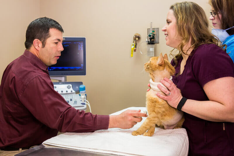
Animal ultrasounds are becoming more readily available in the veterinary field. So, it can be hard to decide who to have perform your pet’s ultrasound. At SCVIM, our Board Certified Internal Medicine Specialists use their expertise as Internists to perform and interpret ultrasounds routinely, and have advanced experience with diseases of the endocrine, gastrointestinal, hematologic, urogenital systems. Their experience and expertise in ultrasound allows them the advantage of seeing the whole picture. We can also use our ultrasound machine to perform ultrasound-guided fine needle aspirates or collect biopsies. These procedures allow us to gather fluid samples or tissues for analysis and to aid in diagnosis of your pet.
In contrast, many other facilities perform an ultrasound but are unable to interpret the images. These images are then sent to a third party (radiologist) for interpretation. In these cases, the radiologist never sees or lays hands on your pet and is limited to the images they are given to make a diagnosis. SCVIM is able to provide a full picture by performing and interpreting our own ultrasounds in-house.
About Veterinary ULTRASOUND
Veterinary ultrasound is a non-invasive procedure that allows two-dimensional visualization of the organ systems within the abdomen. It is more sensitive that radiographs which are two-dimensional by allowing visualization of the internal structures of the organs. Patients are usually given a close haircut and are positioned on their side or back for the duration of the procedure. During an ultrasound examination of the abdomen, we are looking for any architectural or anatomical changes to the organs, evidence of masses or tumors, enlarged lymph nodes, the presence of fluid or gas within the abdominal cavity, evidence of obstruction of tubular structures such as the common bile duct or ureters, stones within the urinary bladder or other parts of the renal system, and signs of changes within the organ systems that may be related to the development of disease processes.
Ancillary testing that may be recommended with abdominal ultrasound:
- Fine needle aspiration with cytology. This procedure involves inserting a needle into the area of interest and collecting a small sample that can be placed on a slide for microscopic evaluation by a board-certified pathologist. The procedure is safe and does not generally require sedation or anesthesia.
- Cystocentesis of the urinary bladder for urine culture. This procedure involves inserting a needle into the urinary bladder to collect a sample of urine. This sample can be evaluated microscopically (urinalysis) or be cultured to evaluate for infection (culture).
- Ultrasound-guided biopsy. This procedure is more involved than a fine needle aspirate. It requires evaluation of the bloods ability to clot normally prior to the procedure and general anesthesia during the procedure. Multiple core biopsy samples are collected and submitted to the pathologist for review and culture. We recommend that pets experiencing biopsy of the liver be hospitalized overnight. There is some risk associated with ultrasound-guided biopsy of the liver, including the risk of anesthesia and hemorrhage from the biopsy site. These risks are uncommon and this procedure is much less invasive than a surgical biopsy, as well as less expensive.
- Abdominocentesis. This procedure is very simple and involves removing fluid that has accumulated in the abdominal cavity. In some instances, only a small volume of fluid is removed for diagnostic purposes. In other cases, larger volumes are removed to make your pet more comfortable. Samples are usually submitted for pathology review and culture.

What Makes an Ultrasound at SCVIM UNIQUE?
Each Ultrasound is performed and interpreted by a SCVIM internist, hands-on with immediate results.
- Our team of 6 internists often consult and collaborate with each other on ultrasounds, with years of clinical feedback (surgical findings and re-evaluations following treatment) to shape and validate our conclusions.
- As the ultrasound evaluation is performed by the Internist, your concerns and your pet’s condition is understood first hand.
- Our ultrasound machines provide enhanced image quality, allowing detection of small and subtle lesions.
Our team practices fear-free techniques, keeping the ultrasound experience for your pet as comfortable as possible.
- We keep your primary care veterinarian informed.
